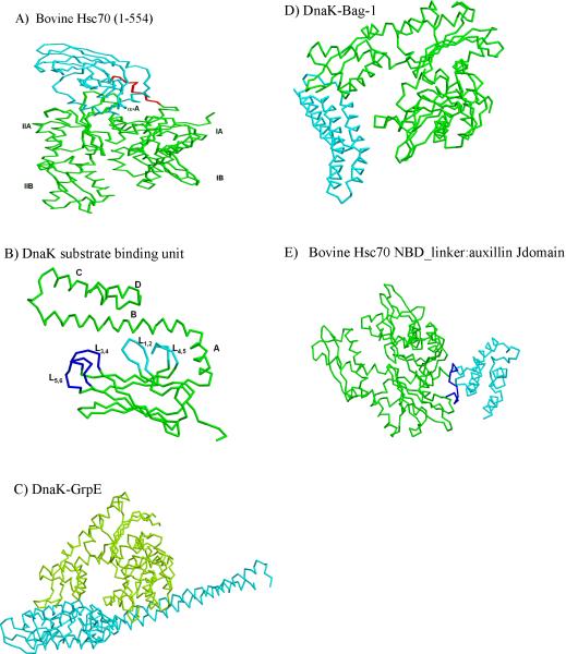
Figure 2.
Structural features of Hsp70 with and without various co-factors. A) Backbone representation of bovine Hsc70 with NBD (green), SBD(cyan) and linker (red) (PDB code 1YUW). B) Backbone representation of substrate binding domain and part of C-terminal domain (389–591) of DnaK (PDB code 1DKY). Inner and outer loops of substrate binding domain are shown in cyan and blue repectively. C) Structure of nucleotide exchange factor GrpE (cyan) bound to ATPase domain (green) of DnaK (PDB code 1DKG). D) Structural representation of the complex of Hsc70 ATPase domain (green) in complex with Bag domain fragment (Cyan) of NEF Bag-1 (PDB code 1HX1). E) Strucutral presentation of auxillin J domain (cyan) bound to bovine Hsc70 NBD (green) containing interdomain linker (blue) (PDB code 2QWO).

