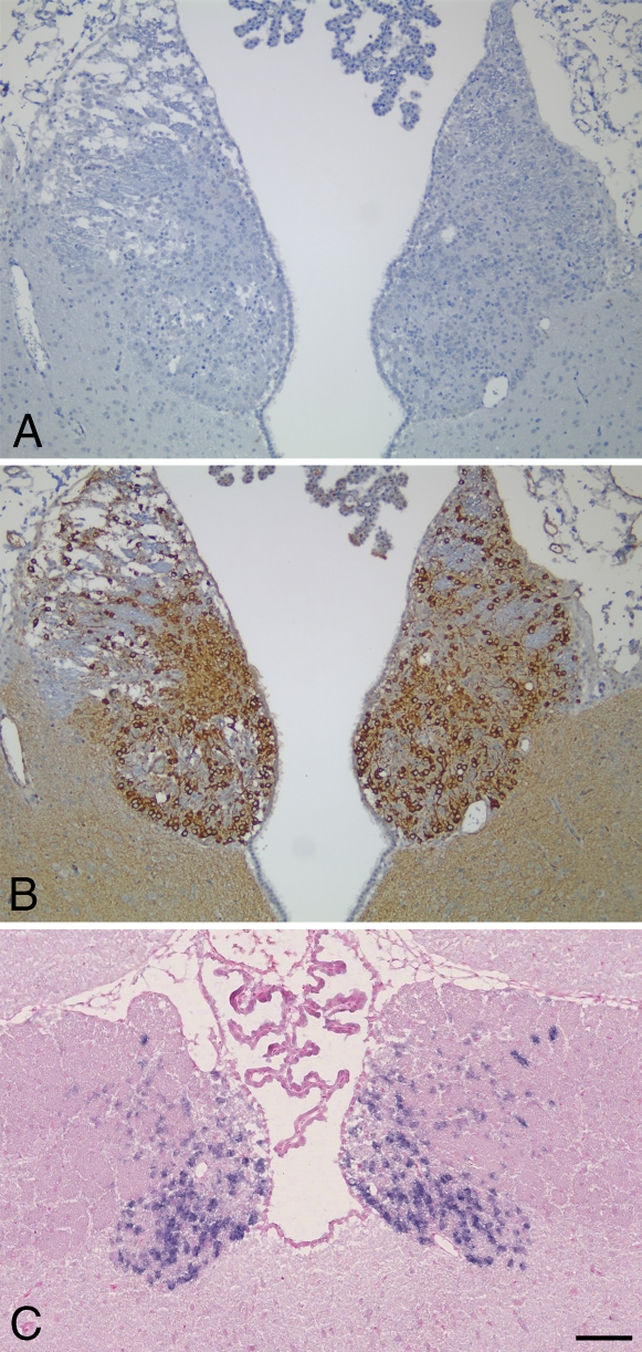
Figure 6.
PDE2A is highly expressed in rat habenula. (A) Coronal section of rat forebrain incubated with anti-PDE2A antibody immunoadsorbed with PDE2A peptide immunogen. (B) Near-adjacent coronal section demonstrating PDE2A immunoperoxidase staining in the rat habenula. (C) Chromogenic in situ hybridization of PDE2A in rat habenula. Blue reaction product is abundant in neurons of the medial habenula. Sections are counterstained with hemotoxylin (A,B) or Nuclear Fast Red (C). Bar = 100 μm.

