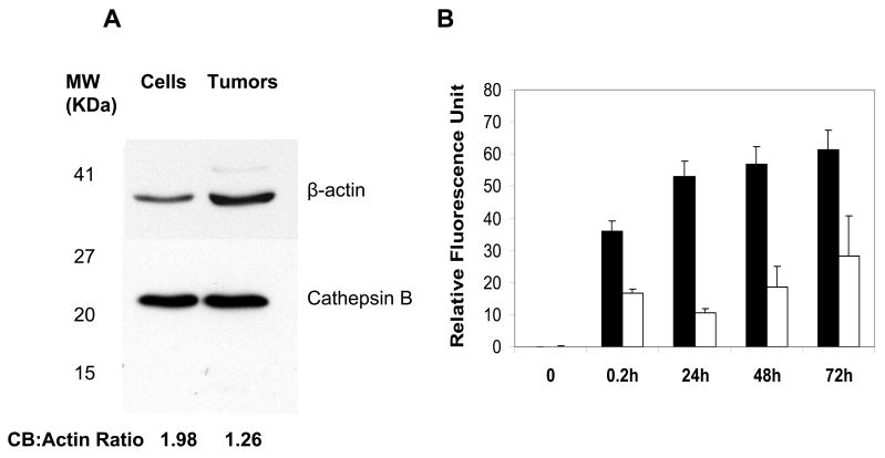
Fig. 5.
Degradation of L-PG-NIR813 in U87/TGL cells. A: Western blot analysis of CB expression in U87/TGL cell lysate and excised U87/TGL tumors. B: In vitro degradation of L-PG-NIR813 in cultured U87/TGL cells as measured by activation of fluorescence signal. The data represent mean ± SD (n = 3). Note the significantly higher signal intensity from 0.2 h to 72 h in U87/TGL cell culture (■) than in fresh culture medium (□) (p < 0.001).

