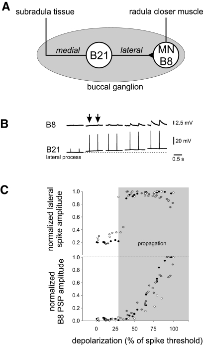
FIG. 1.
A: schematic of the preparation. Mechanical stimulation of the subradula tissue evokes spikes, which travel along the medial process, across the central soma, and into the lateral branch where the main contacts between B21 and follower motor neuron B8 are located. B: effects of depolarization on B21–B8 transmission. Simultaneous recordings from B21's lateral process and B8 during peripheral activation of B21. The dashed line under the lateral process trace indicates the resting membrane potential. The 1st 2 responses were evoked at resting membrane potential. Thereafter B21 was progressively depolarized by current injection into the soma (trace not shown). In each case, 2 responses are shown at each membrane potential. When B21 was peripherally activated at resting potential, spikes did not actively propagate to the lateral process, and PSPs in B8 were virtually nonexistent. As B21 was depolarized, the spike propagation failure was relieved, but initially PSPs in B8 were virtually undetectable (arrows). With further depolarization, PSP amplitude progressively increased. C: summary of data for the experiment shown in B from 4 animals, each plotted with a different symbol. y axis data have been normalized to maximum spike height or peak PSP amplitude. X axis data have been normalized to the amount of depolarization that was needed to evoke spikes centrally. The shaded region indicates the approximate point at which spike propagation to the lateral process begins. Note that the B8 PSPs are still virtually undetectable at this point. Also note the graded, progressive increase in PSP amplitude as the holding potential was progressively increased.

