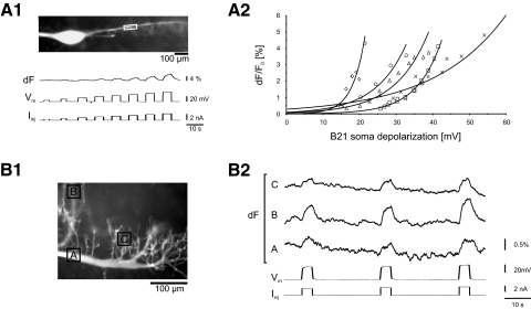
FIG. 2.
Subthreshold depolarizations increase the intracellular calcium concentration in B21. A1, photo: low-power (×10) image of a neuron B21 after injection of the Ca2+ indicator dye Calcium Orange. The box on the lateral process marks where optical measurements were made in the recordings shown below. Traces: subthreshold current pulses of progressively increasing amplitudes were injected into the soma of B21 (Iinj). These pulses induced changes in membrane potential (Vm) and graded increases in calcium fluorescence (dF). A2: group data and fitted exponential functions from 5 experiments like the 1 shown in A1. Note the graded increase in dF as pulse amplitude was increased. B1: higher (×40)-power image of a B21 neuron injected with Calcium Orange. The boxes mark the regions imaged in B2. Subthreshold current pulses were injected into the soma of B21 (Iinj), which induced changes in membrane potential (Vm) and increases in calcium fluorescence (dF).

