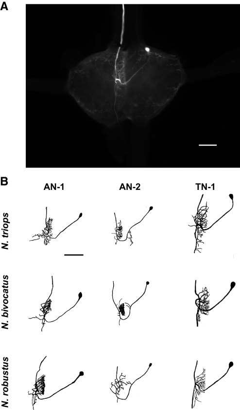
FIG. 1.
A: photo of TN-1 filled with Lucifer yellow in Neoconocephalus triops to show the location of the auditory interneurons within the prothoracic ganglion. The cell bodies of all 3 auditory neurons with ascending axons are found within the same region as the TN-1 cell body in the photo for all 3 species. The dendritic field for the TN-1 shown also represents the area where auditory receptor cells terminate and form the auditory neuropile. The axon ascends to the supraesophageal ganglion (SOG) in the soma–contralateral connective. B: morphology of the 3 primary auditory interneurons that have ascending axons (AN-1, AN-2, and TN-1) for the 3 Neoconocephalus species tested. Auditory neurons were drawn from digital stacked images to show the similarity in morphology of the 3 interneurons across the 3 species. The TN-1 neuron for N. triops is the same as in A. Scale bars: 150 μm.

