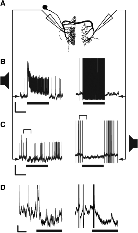
FIG. 9.
A: morphology of the local omega neuron in N. triops. Omega morphology was similar in the other 2 species. The dendritic processes are located on the soma-ipsilateral, whereas the axonal processes are on the soma-contralateral side. B: dendritic (left) and axonal (right) responses to conspecific call stimuli presented from the soma-ipsilateral (preferred) side. Axonal responses were 60 mV, but truncated at 40 mV in the figure. Scale bars: 500 ms, 10 mV. C: dendritic and axonal responses to the same stimulus presented from the soma-contralateral side. Axonal responses truncated at 40 mV. Brackets indicated portions of traces expanded in D. Scale bars: 500 ms, 10 mV. D: traces in C expanded to better show the hyperpolarization in both axonal and dendritic recordings. AP amplitudes are truncated. Black bar represents the stimulus. Scale bars: 100 ms, 2 mV.

