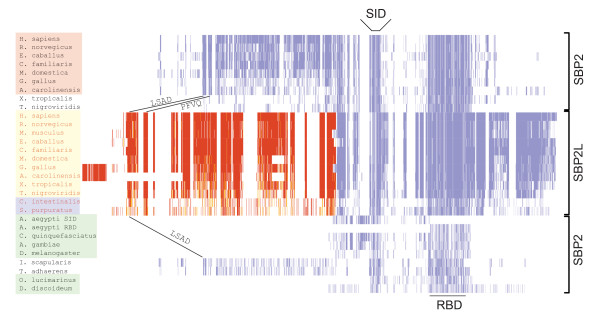
Figure 1.
SECIS binding protein topology. Alignment of SBP2 and SBP2L across the species indicated generated with the MUSCLE module in Geneious (Biomatters Ltd). The N and C-terminal portions of SBP2 and SBP2L were independently aligned. Residues were colored using JalView based on BLOSUM62 score [[37]; darker colors indicate higher similarity]. The SBP2L N-terminal sequence is red to denote that it is a separate alignment from that for SBP2. Globally conserved motifs motifs include the Sec incorporation domain (SID), the RNA binding domain (RBD), LSAD15-26 and PFVQ44-56. Shading of species names is used for the identification of SBP2 classes as described in the text.

