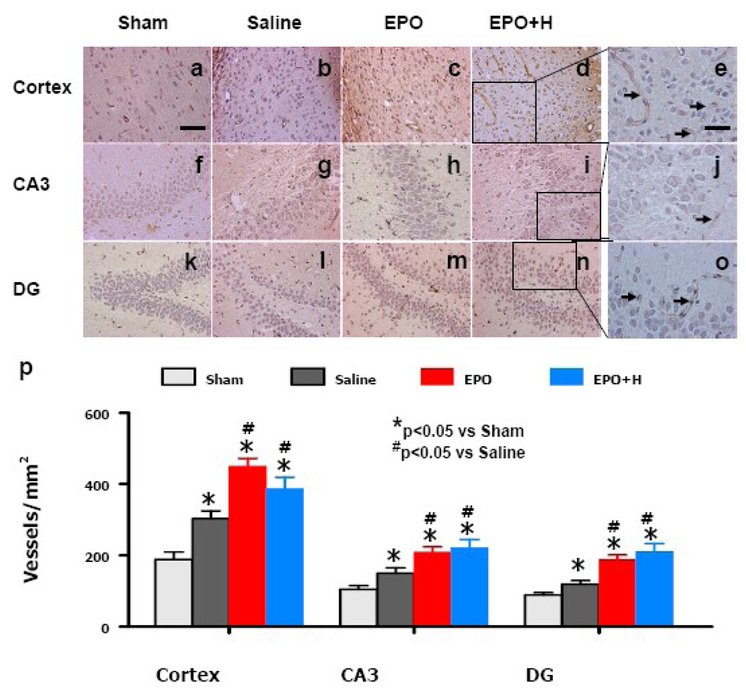
Fig. 4.
Effect of EPO on vWF-staining vascular structure in the ipsilateral cortex, DG and CA3 region 35 days after TBI. TBI alone (b, g, i) significantly increased the vascular density in these regions compared to sham controls (P<0.05). EPO treatment (EPO and EPO+H) further enhanced angiogenesis after TBI compared to saline groups (P<0.05). Hemodilution did not affect vascular density compared to EPO alone group. The density of vWF-stained vasculature is shown in (p). Data represent mean ± SD. * P<0.05 vs. Sham. #P<0.05 vs. the saline group. Data represent mean ± SD. N = 6 (Sham); 6 (Saline); 6 (EPO); 7 (EPO+H). Scale bar = 50 µm (a); 25 µm (e).

