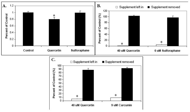
Fig. 7.
The effects of in vivo phytochemical supplementation on mouse splenocyte proliferation, and the retention of phytochemical supplement effect “recall”. (A) BALB/c mice were fed a powdered diet of AIN-93M with or without quercetin (0.25 w/w) or sulforaphane (0.05 w/w) for 10 days. After sacrifice, splenocytes were isolated from spleens, pretreated with the indicated phytochemical supplements at various concentrations for 30 minutes, stimulated with anti-CD3 plus anti-CD28, and proliferation measured by tritiated thymidine incorporation. Data is expressed as means +/− SEM (N=4). (B) and (C) Retention of phytochemical supplement recall. (B) Mouse splenocytes were supplemented with 40 uM quercetin or 6 uM sulforaphane for 30 minutes, centrifuged, washed, and split prior to plating. Half of these culture wells were then resupplemented with 40 uM quercetin and 6 uM sulforaphane, and the other half with solvent (DMSO) only. Control (unsupplemented) cultures were also included. Cells were then stimulated with anti-CD3 plus anti-CD28 and proliferation measured by tritiated thymidine incorporation. (C) Human T-lymphocytes were supplemented with 40 uM quercetin or 9 uM curcumin for 30 minutes, centrifuged, washed, and split prior to plating. Half of these culture wells were then resupplemented with 40 uM quercetin and 9 uM curcumin, and the other half with solvent (DMSO) only. Control (unsupplemented) cultures were also included. Cells were then stimulated with anti-CD3 plus anti-CD28 and proliferation measured by tritiated thymidine incorporation. Statistically significant differences between control and treated samples (A) and between supplement left in versus supplement removed (B and C) were assessed by the Kruskal-Wallis test. *P < .05.

