
Figure 5.
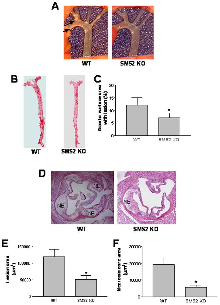
SMS2−/−→LDLr−/− mice demonstrated significantly decreased atherosclerotic lesion size and necrotic area. Panel A, mice were euthanized, and the aortas dissected and photographed. This set of pictures is representative of 9 sets. Atherosclerotic lesions are indicated by arrows; Panel B, Oil Red O staining for whole aorta (en face assay); Panel C, quantitative display of en face assay; Panel D, hematoxylin and eosin staining for proximal aorta (root assay) (NE: necrotic core area); Panel E, quantitative display of root assay; Panel F, quantitative display of necrotic core area. Values are mean ± SD, N=9. *P<0.01.