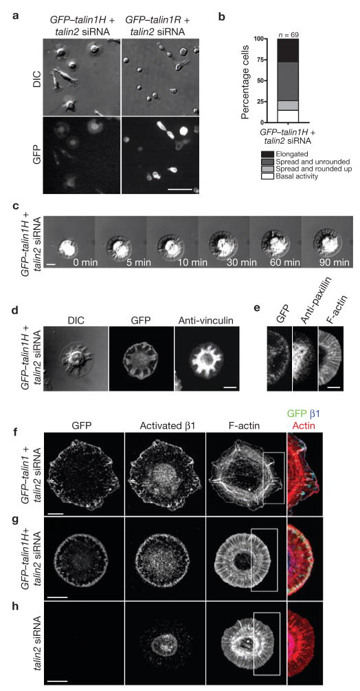
Figure 3.
Talin1 head activates β1 integrin but does not rescue focal adhesion formation. (a) The spread phase on fibronectin was prolonged in cells expressing the talin1 head (talin1H) but not the talin1 rod (talin1R). Talin1−/− cells co-transfected with GFP-talin1H and talin2 siRNA or with GFP-talin1R and talin2 siRNA were plated on fibronectin for 90 min before fixation. (b) Summary of early spreading behaviour of talin1H-expressing cells. (c) Sequential time-lapse images of the talin1H-expressing cell spreading on fibronectin (Supplementary Information, Movie 4). (d, e) Focal adhesion formation was defective in talin1H-expressing cells. 90 min after plating, cells were fixed and stained for vinculin (d) and paxillin (e). (f–h) Talin, through its head domain, activated and colocalized with β1 integrin at the leading edge of spread cells. GFP–talin1-restored cells (f), GFP–talin1H-expressing cells (g) and talin-deficient cells (h) were plated on fibronectin for 20 min, 90 min and 20 min, respectively before fixation. Scale bars are 50 μm (a), 5 μm (e) and 10 μm (c, d, f–h).

