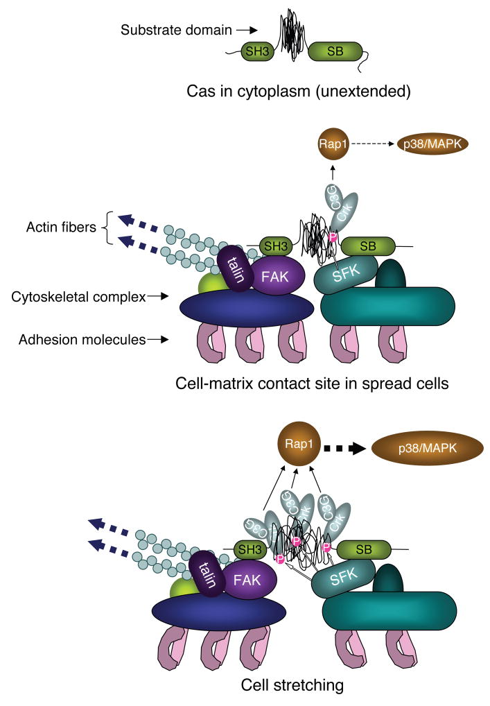
Figure 6. Model of Extension of Cas and Signaling at Cell-matrix Contact Sites.
The top and middle panels represent a Cas molecule with unextended configuration of substrate domain in the cytoplasm and with moderate extension of substrate domain at the cell-matrix contact site of spread cells, respectively. The bottom panel represents the extension-dependent phosphorylation of Cas substrate domain by SFK and enhancement of its downstream signaling. SH3 and SB represent the SH3 and the Src-binding domains of Cas, respectively.

