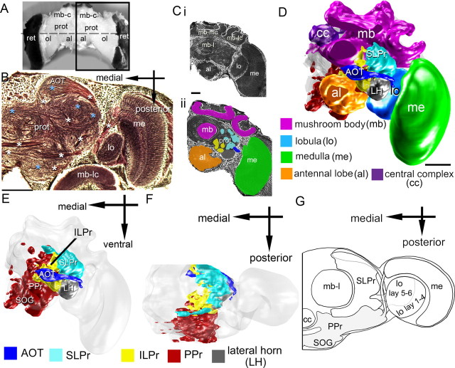
Figure 1.
The bee brain and the lateral protocerebrum. A, Whole bumblebee brain viewed frontally. Several structures are noted, including the optic lobes (ol), mushroom body calyces (mb-c), antennal lobes (al), retina (ret), and protocerebrum (prot). B, Bodian-stained horizontal section at the level of the dashed line in A. The staining delineates the lobula (lo), medulla (me), the mushroom body lateral calyx (mb-lc), and the protocerebrum. Glomeruli in the protocerebrum (prot) are indicated by light blue asterisks, axonal tracts by white asterisks. Ci, Frontal section of half of the bumblebee brain, Bodian stained. The staining delineates the antennal lobes, the lobula, medulla, the mushroom body lateral calyx, the mushroom body medial calyx (mb-mc), and the mushroom body lobe (mb-l). Cii, Color-coded areas used to reconstruct the brain: the medulla (green), antennal lobes (orange), and protocerebral areas comprising glomeruli (cyan and yellow). D, Three-dimensional reconstruction of major components of the bumblebee brain, including the antennal lobes (orange), lobula (light blue), medulla (green), central complex (cc, purple), mushroom bodies (mb, magenta), LH (gray), and SLPr (cyan). E, Reconstructions of brain areas as in D along with other brain areas removed to reveal protocerebral components: LH (gray), SLPr (cyan), ILPr (yellow), AOT (blue), PPr (red), and subesophageal ganglion (SOG). F, The same brain reconstruction rotated for a dorsal view. G, A schematic of a dorsal view of the same brain areas, with a view of the SLPr, PPr, optic lobes, and other brain areas listed above. Scale bar, 100 μm.

