
Figure 3.
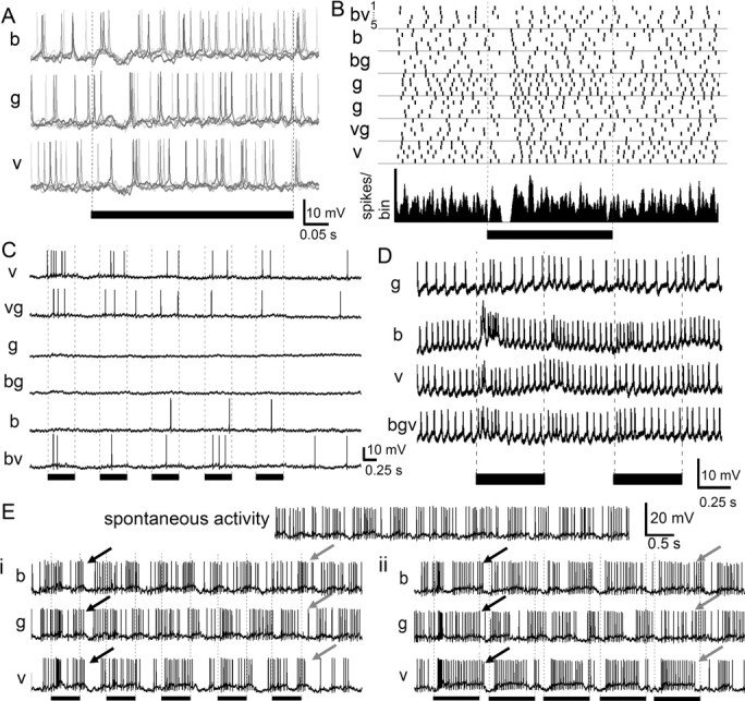
Color responses of protocerebral neurons. A, Protocerebral neuron responding with phasic “on” inhibition for all three colors [blue (b), green (g), and violet (v)] and across five trials (traces from each trial are overlaid and colored in gray). B, Raster plot (top; five trials per color) and peristimulus time histogram [bottom; Gaussian-smoothed (half-width = 5 ms); bin width = 1 ms] of responses of the same neuron as in A, revealing its phasic inhibition by light, regardless of color and color combinations (bv, vg, bg). C, Protocerebral neuron responding when violet light was present in the stimulus but not or only weakly to the other colors. D, Protocerebral neuron producing a burst of activity in response to the first light flash of blue, with a decreased response to violet and green. Simultaneous presentation of all three colors results in reduced activity compared with blue alone, suggesting that other colors inhibit the blue response. E, Stimulus entrainment. This protocerebral neuron exhibited a spontaneous rhythm of activity which persisted throughout the recording. Ei, The neuron shifts its bursting activity over time to match the rhythm of the violet light flashes. Green or blue light had a much weaker entrainment effect (black arrows: end of first stimulus; gray arrows: end of fifth stimulus). Eii, The stimulus entrainment response was also observed if the duration of the light flash was lengthened. In all cases, the black bars indicate the duration of the light flash. Stimulus markers: 500 ms in A–E, 840 ms in F.