Figure 5.
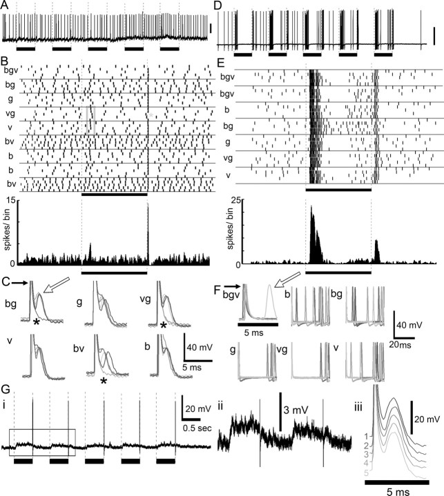
Temporal response properties of protocerebral neurons. A, The recording trace during a series of five light flashes does not exhibit a significant change in spike rate, but when the spike times were aligned across trials (raster in B), there was remarkable precision at the offset of the light flash also seen in the peristimulus time histogram (bin width = 1 ms, smoothed with a Gaussian filter, half-width = 5 ms). The gray box outlines instances of precise responses to the onset of the stimulus. C, Extremely well timed spikes occurred after the light flash regardless of color (white arrow). Five individual trials are overlaid; the large initial spike (black arrow) represents electrical noise produced by the relay switch. In some cases, the neuron fails to respond to the end of the light flash, as indicated by the gray asterisk in B and the black asterisks in C. D, Lobula neuron with pronounced “on” and “off” response but without a precise spike time response, demonstrating that the precise spike timing in A–C was not an artifact of the relay switch noise. E, This neuron (same as in D and F) did not exhibit precisely aligned spikes across trials. F, The spikes were not well timed with the offset of the stimulus (superimposed noise of relay switch; white arrow, action potential spike of the neuron). Gi, Another protocerebral neuron shows tonic depolarization for the duration of the light flash (Gii; box in Gi) and a single highly precise spike after each of a series of five blue light flashes. Giii, All five trials aligned at the end of the light flash (membrane potentials staggered to view each trial); note the highly precise temporal alignment of spikes.

