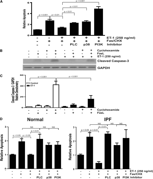
Figure 4.
ET-1–mediated protection from apoptosis is dependent on p38 MAPK and PI3K/AKT. (A–C) IMR-90 fibroblasts in a 96-well plate were treated with the combination of FasL (250 ng/ml) and cycloheximide (500 ng/ml) in the presence/absence of ET-1 (250 ng/ml) with/without inhibitors of PLC (U73122, 5 mM), p38 MAPK (SB203580, 6 μM), or PI3K (Wortmannin, 50 nM) for 16 hours. Apoptosis was assessed by (A) ELISA for ssDNA (four replicates per condition and three separate experiments) and (B) Western immunoblotting for cleaved caspase-3. The membrane in (B) was stripped and probed for GAPDH. (C) Densitometric analysis of cleaved caspase-3/GAPDH ratios from three independent experiments. (D) Similarly treated primary normal lung fibroblasts (left panel) and IPF fibroblasts (right panel) were assessed for apoptosis by ELISA for ssDNA (n = 3 normal lung fibroblast lines and 3 IPF fibroblast lines).

