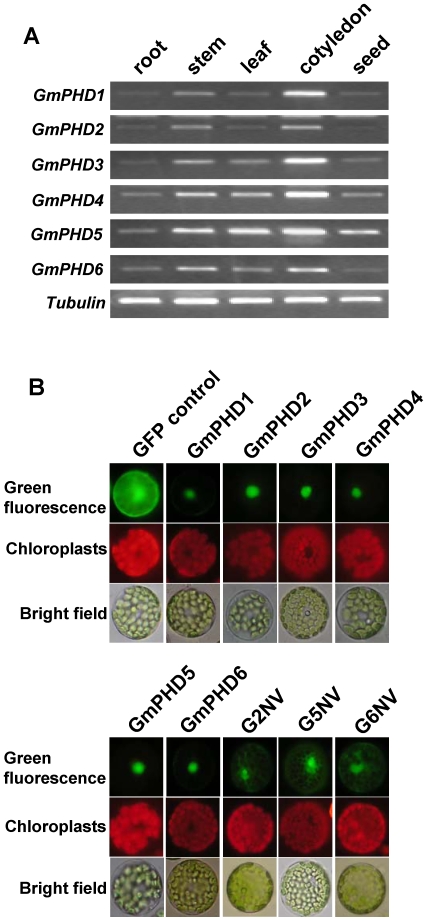
Figure 4. Organ-specific expression and subcellular localization of the GmPHDs.
(A) Expression of GmPHDs in different organs of soybean plants revealed by RT-PCR. A Tubulin fragment was amplified as an internal control. (B) Subcellular localization of GmPHD proteins in Arabidopsis protoplasts as revealed by green fluorescence of. GmPHD-GFP fusions or GFP control. For each panel, the photographs were taken in the dark field for green fluorescence (upper), for red fluorescence indicating chloroplasts (middle), and in the bright light for the morphology of the cells (lower). G2NV: the NV domain of GmPHD2; G5NV: the NV domain of GmPHD5; G6NV: the NV domain of GmPHD6.

