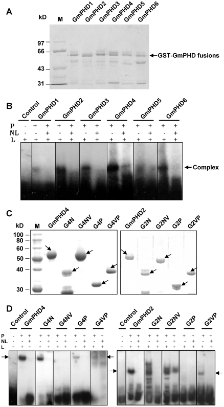
Figure 7. DNA-binding specificity of the GmPHD proteins.
(A) Coomassie blue staining of the six GST-GmPHD fusion proteins on SDS/PAGE. Arrow indicates the fusion proteins. Lower bands probably represent the degradation products. (B) Gel shift assay of the six GmPHD proteins. GmPHD proteins (P) were incubated with a radiolabeled probe containing 5 X GTGGAG (L), in the presence (+) or absence (−) of unlabeled probes (NL) in ten-fold excess. Arrow indicates position of the protein/DNA complexes. (C) Coomassie blue staining of various domains of the GmPHD4 and GmPHD2. Arrows indicate the corresponding proteins. (D) Gel shift assay of the GmPHD4, GmPHD2 and their domains. Others are as in (B). Arrows indicate positions of the protein/DNA complexes.

