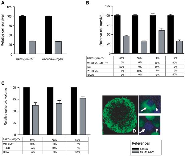
Figure 4.
In vitro sensitivity to ganciclovir of heterotypic cell cultures and spheroids containing stromal cells expressing TK. A, ganciclovir sensitivity of BAEC and WI-38 VA cells expressing TK. In all cases, ganciclovir was added for 5 d, the day after cells were seeded on the plates; data were obtained by the 3-(4,5-dimethylthiazol-2-yl)-2,5-diphenyltetrazolium bromide assay. B, ganciclovir sensitivity of cells monolayers containing BAEC-(Δ10)-TK or WI-38 VA-(Δ10)-TK cells mixed with A375N cells. C, ganciclovir sensitivity of heterotypic spheroids made of BAEC-(Δ10)-TK cells mixed with different malignant cell types. D, immunofluorescence of a heterotypic spheroid made of Mel-EGFP and BAEC-(Δ10)-TK cells showingthe presence of Mel-EGFP green fluorescent cells at the outer part. E, combined immunofluorescence of EGFP and proliferatingcell nuclear antigen on a spheroid made of Mel-EGFP and BAEC-(Δ10)-TK cells. A representative Mel-EGFP cell (arrow) showing a blue nucleus (corresponding to positive proliferating cell nuclear antigen staining) and green cytoplasm. F, similar to E but showinga potential BAEC-(Δ10)-TK cell (arrow) with a blue nucleus and no green fluorescence in the cytoplasm.

