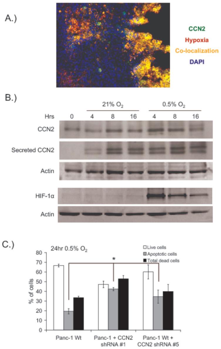
Figure 5. CCN2 expression and secretion is increased by hypoxia and protects pancreatic tumor cells from hypoxia-induced apoptosis.
(A) Pimonidazole (Hypoxyprobe-1) was administered 90 minutes prior to orthotopic Panc-1 tumor excision, and frozen tumor sections were analyzed for pimonidazole (red) and CCN2 (green). Areas of co-localization are indicated (yellow), and nuclei are stained with DAPI (blue).
(B) Western blots of CCN2 and HIF-1α in Panc-1 cells incubated at 21% or 0.5% oxygen for the indicated periods of time before collection of cell lysate and conditioned media. Secreted CCN2 was obtained by contacting conditioned media with heparin sepharosecoated beads prior to loading. Actin in the cell lysate was used as a loading control.
(C) Flow cytometric quantification of wild-type Panc-1 cells or Panc-1 shRNA-expressing clones exposed to 0.5% O2 for 24hr. Apoptotic cells were measured by annexin-V staining and quantified by flow cytometry. *p<0.05 relative to control cells.

