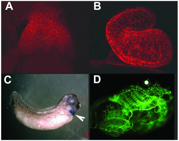
Figure 1.
Morphological and spatial analysis of cardiovascular development. Panels A and B show whole mount confocal images of the Xenopus heart at stage 28 and 35. Heart tissue was detected by an anti-tropomyosin antibody. Panel C shows the in situ hybridization detection of Nkx2-5 mRNA. Cardiac regions (indicated by the arrowhead) expressing Nkx2-5 appear purple. Panel D shows an embryo transgenic for the flk promoter driving a GFP reporter. This transgenic embryo expresses GFP in the vascular system and was used to delineate the important regulatory elements of the flk promoter.

