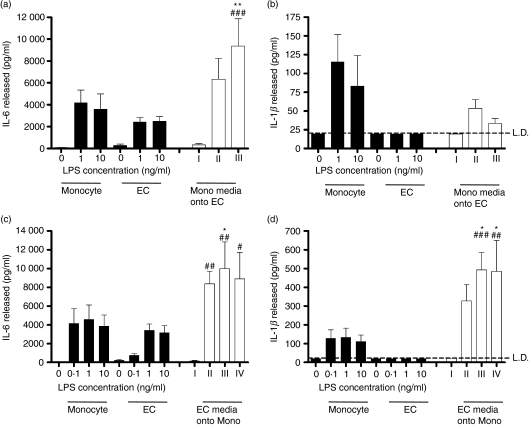
Figure 3.
Endothelial cells (EC) are a source of interleukin-6 (IL-6) but not IL-1β in the cocultures, whereas as monocytes are a source of both cytokines. Monocytes or EC were cultured in isolation, at the same densities and conditions as used in cocultures. Filled bars show IL-6 and IL-1β production after direct activation of monocytes or EC by the indicated concentrations of lipopolysaccharide (LPS). In open bars, supernatants from LPS-treated monocytes were transferred onto fresh EC, or vice versa. (a, b) Bar I indicates the effects of addition of supernatants (diluted one in two in fresh media) from buffer-treated monocytes on EC. Bars II and III indicate the addition of media from monocytes treated with 1 or 10 ng/ml LPS, respectively, similarly diluted 1 : 2 in fresh media. (a) Release of IL-6 from EC treated with media from LPS-stimulated monocytes, and (b) media from LPS-stimulated monocytes was unable to induce the release of IL-1β from EC. (c, d) Bar I indicates the addition of supernatants (diluted one in two in fresh media) from buffer-treated EC onto monocytes; bars II, III and IV indicate the addition of media from EC treated with 0·1, 1 or 10 ng/ml LPS, respectively, similarly diluted 1 : 2 in fresh media. (c, d) Media from LPS-treated EC enhanced the release of both IL-6 and IL-1β from monocytes. Data are ± SEM (n = 4–8), each experiment using cells from independent donors. **P < 0·01 and *P < 0·05 (comparing the indicated bars with cytokine levels generated by monocytes alone). ###P < 0·001, ##P < 0·01 and #P < 0·05 (comparing the indicated bars with cytokine levels generated by EC alone). Data were analysed using two-way anova with Bonferroni’s post-test. LD indicates the limit of detection of the enzyme-linked immunosorbent assay.

