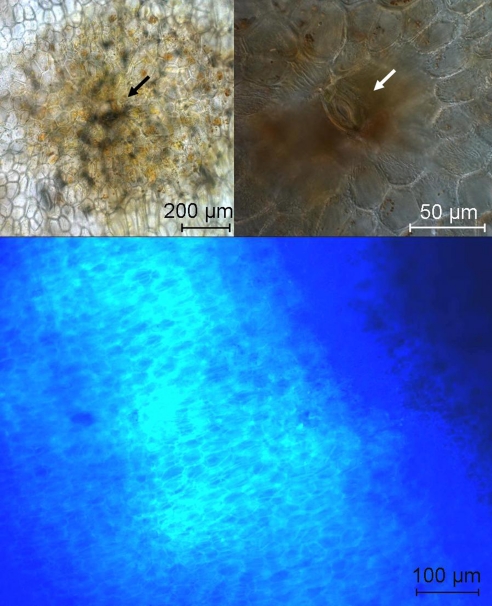
Fig. 5.
Senescence associated dark spots originate from stomata. (Top Left) Bright field microscopic images of dark spot on the surface of a yellow banana peel, stoma in the center (see arrow). (Top Right) Differential interference contrast (DIC) image with higher magnification, guard cells of the stomata are clearly visible in the center of the dark spot (see arrow). (Bottom) Fluorescence microscopic image of the transition zone between a dark spot and its yellow surroundings on the surface of a banana peel (all images depict surface sections of the epidermis and subepidermal parenchyma tissue, for experimental details, see Materials and Methods).

