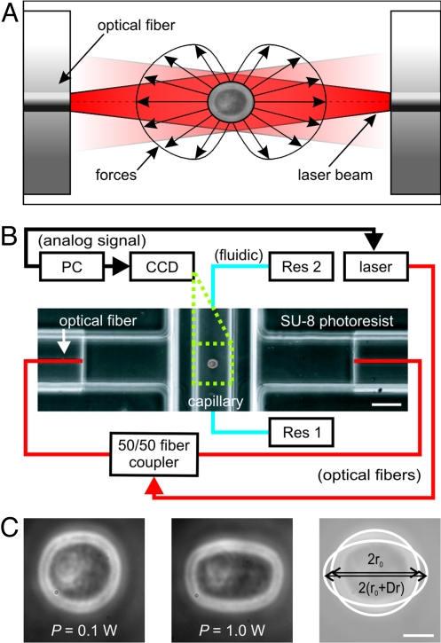
Fig. 1.
Principle and setup of a microfluidic optical stretcher (μOS). (A) Two-counter propagating NIR laser beams (λ = 1,064 nm) emanating from the cores of single-mode optical fibers are used to trap (P = 0.1 W each beam) and deform (P = 1 W each beam) single cells. Cells are deformed by the forces arising from the momentum transfer of light to the surface of the cell due to the change in refractive index. (B) Experimental setup. The flow of the cell suspension between two reservoirs (Res 1 and Res 2) is controlled by a hydrostatic pressure differential. The phase contrast image shows the microfluidic flowchamber with a cell trapped inside the glass microcapillary. (C) Phase-contrast images of APL cell being optically trapped (left) and stretched (middle) in the μOS. Optically induced surface stress causes a deformation of the cell along the optical axis. (Right) overlay of the two other images and definition of the strain, γ(t) = Δr(t)/r0. (Scale bar, 5 μm.)

