Fig. 2.
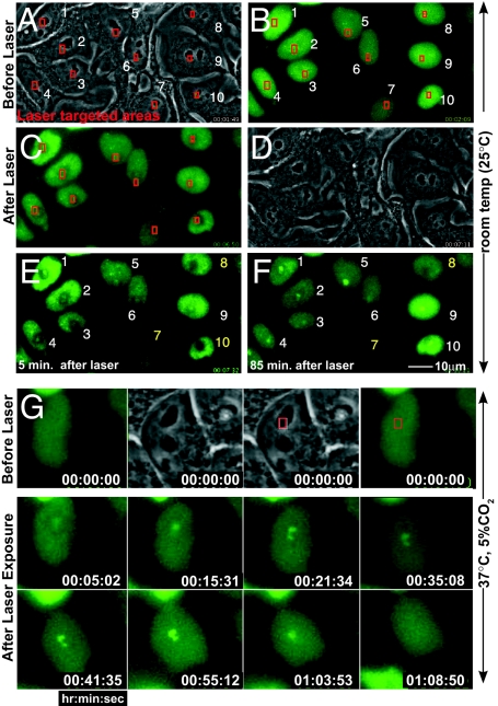
Rapid GFP-CENP-A accumulation at sites of DNA damage. (A–F) GFP-CENP-A cells before and after laser targeting at 25 °C. Phase contrast (A) before and (D) 5 min. after targeting the areas boxed in red. Epifluorescence images of GFP-CENP-A, immediately (B) before and (C) 4 or (E) 5 min. after initiating laser exposure. In most cells (numbered in white), GFP-CENP-A accumulated at the sites of targeting. [Cells #7, 8 and 10 (numbered in yellow) were bleached during laser targeting,] (F) Within 85 min, GFP-CENP-A formed foci within targeted regions. (G) Laser targeting as in (A–F), maintained at 37 °C after targeting: a CENP-A focus appears within ≈5 min after laser exposure, reaches its peak intensity ≈1 h after laser exposure, and then disappears approximately 15 min later. Timestamp represents hours:minutes:seconds.

