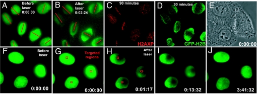
Fig. 4.
Histone H2B never accumulates in areas of laser-induced DNA damage. (A–D) Epifluorescence images of HeLa cells stably expressing YFP-H2B (A) before and (B–D) after laser exposure (red lines). (C) γ-H2AX and (D) YFP-H2B 90 min. after laser exposure. (E) Phase contrast image of HeLa cells stably expressing YFP-H2B before laser targeting. (F–J) YFP-H2B epifluorescence of the cells in (E) and (F and G) before or (H–J) after laser targeting. Red squares in (G and H) denote laser targeted areas. Timestamp is hours:minutes:seconds.

