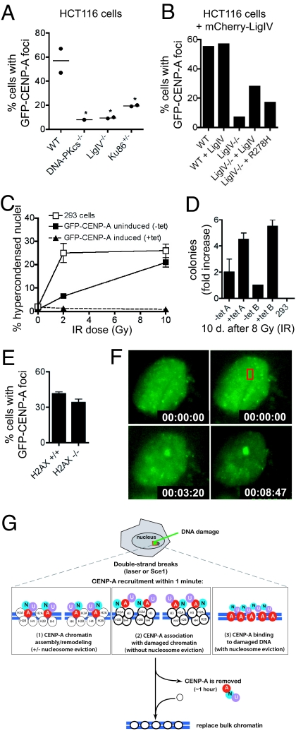
Fig. 6.
CENP-A in function in DNA repair independent of H2AX, and possible models for CENP-A assembly at sites of DNA repair. (A) HCT116 cells generated by targeted disruption of NHEJ genes were transiently transfected to express GFP-CENP-A; frequencies of focus formation were measured after laser exposure. [WT (n = 47 cells); DNA-PKcs−/− (n = 39 cells); LigIV−/− (n = 57 cells); Ku86+/− (n = 38 cells); P = 0.0202 (one-way ANOVA)]. (B) Frequencies of GFP-CENP-A recruitment to sites of laser-induced DNA damage in Ligase IV−/− cells transfected to express mCherry-LigaseIV or mCherry-LigaseIV-R278H. (C) Percentages of cell death following 0 Gy, 2 Gy, and 10 Gy irradiation (done in triplicate) in cells with a stably integrated, GFP-CENP-A gene, either with or without tetracyline-mediated gene induction. Cell death was scored by small, bright nuclei [using Hoechst] and quantified 24 h later by automated microscopy (n = 2,500–5,000 cells per sample). Error bars show the standard devia tion. (D) Cell survival in a colony formation assay after 8 Gy irradiation of parental 293 cells or inducible GFP-CENP-A cells. (E) Frequency of (transiently transfected) GFP-mCENP-A recruitment to sites of laser-induced DNA damage in wild-type and H2AX null MEFs. (F) GFP-mCENP-A recruitment to focal DNA damage (red box) in an H2AX null cell. (G) Model for CENP-A recruitment to sites of DNA damage. Laser-mediated damage or Sce1 cleavage creates DNA double-strand breaks. CENP-A, CENP-N and CENP-U are recruited within 1–2 min. Three possible modes of CENP-A binding are shown (see Discussion).

