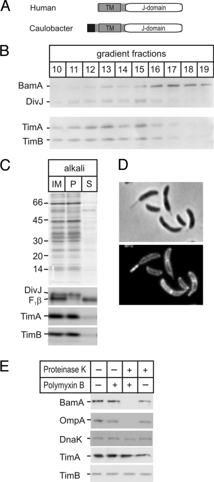
Fig. 2.
Location and topology of TimA and TimB. (A) Domain structure of Tim14 (41, 42) and TimB. (Black, signal sequence; TM, transmembrane domain.) The “J-domain” (31) that interacts with Hsp70 is shown (detailed sequence analysis provided in Fig. S2). (B) Membranes were fractionated on sucrose gradient and analyzed by SDS/PAGE and immunoblotting. (C) Inner membrane vesicles (IM) were extracted with alkali and the pellet (P) and supernatant (S) fractions analyzed by Coomassie staining (Upper) and immunoblots for TimA, TimB, the integral membrane protein DivJ, and peripheral membrane protein F1β. (D) Fluorescence microscopy of the OmpA-mCherry strain shows the periplasmic location of the protein. (E) Caulobacter cells were incubated without (−) or with (+) polymyxin B and proteinase K as indicated and then analyzed by SDS/PAGE and immunoblotting.

