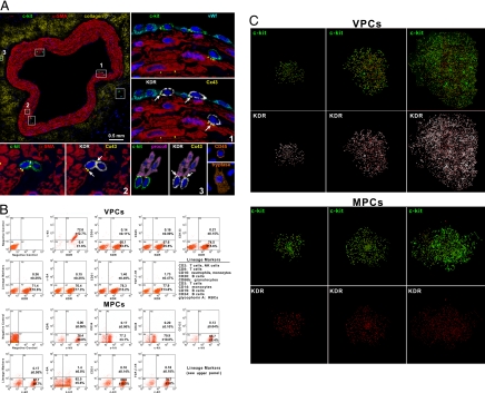
Fig. 1.
Localization and properties of vascular progenitor cells (VPCs) and myocyte progenitor cells (MPCs). (A) Cross-section of human epicardial coronary artery composed of several layers of smooth muscle cells (SMCs) [α-smooth-muscle-actin (α-SMA); red]. The c-kit-positive cells (green) are included in six rectangles. Three of the six rectangles are shown at a higher magnification in the adjacent panels. The c-kit-positive cells express KDR (white). Connexin 43 (Cx43; yellow dots; arrows) is seen between c-kit-KDR-positive cells and endothelial cells (von Willebrand factor; bright blue), SMCs (α-SMA; red), and adventitial fibroblasts (procollagen, procoll; magenta). The c-kit-KDR-positive cells are negative for CD45 and tryptase, excluding mast cells. Insets: Positive controls. See Fig. S1A in SI Appendix for the other three areas. (B) FACS of VPCs and MPCs. Negative controls were used for all epitopes. Two examples corresponding to isotype-matched antibodies for c-kit and KDR are shown. (C) Clones derived from single sorted VPCs and MPCs.

