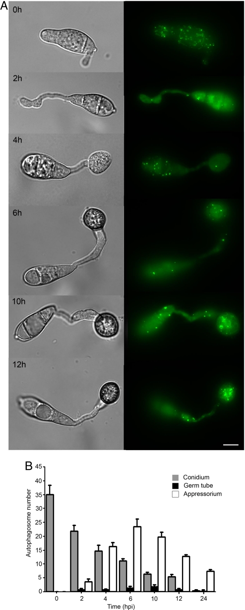
Fig. 1.
Cellular localization of autophagosomes during infection-related development of M. oryzae. (A) Conidia were harvested from a Guy-11 transformant expressing a GFP:MoATG8 gene fusion, inoculated onto glass coverslips, and observed by epifluorescence microscopy at the times indicated (Scale bar, 10 μm.). (B) Bar chart showing mean autophagosome numbers present in conidium, germ tube and appressorium at 0 h, 2 h, 4 h, 6 h, 10 h, and 12 h after inoculation (error bars indicate ± 2 SE).

