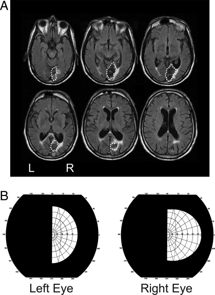
Fig. 1.
Lesion location and visual field testing in patient CB. (A) FLAIR MRI images (5-mm axial slices) of patient CB's lesion acquired 3 years post-stroke. The lesion is largely restricted to the right occipital region and the optic radiations. The dotted lines indicate the boundaries of his lesion. (B) Results of Goldman perimetry conducted at the time of testing (3.5 years post-stroke) by a neuro-ophthalmologist. Testing was conducted using III 4 sized targets, which are commonly used to assess visual field defects after stroke. Importantly, there is no evidence of any macular or temporal crescent sparing in the left (blind) field.

