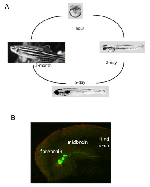
Figure 1.
The zebrafish life cycle (A) and the developing brain (B, lateral view of ~ 1-day old embryonic head region with the dorsal side up), with the visualization of dopaminergic (anti-TH, green) and serotonergic (anti-serotonin, red) neurons. Images in (A) are form ZFIN.org. Dopaminergic and serotonergic neurons are visualized through antibody labeling of tyrosine hydroxylase and 5HT respectively.

