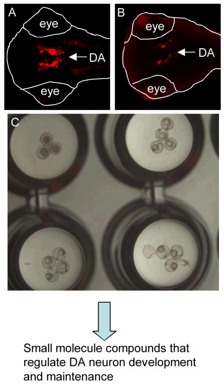
Figure 2.
Identification of small molecule compounds that can regulate DA neuron development and maintenance. DA neurons are visualized through tyrosine hydroxylase immunostaining in normal (A) and MPTP-treated (B) embryos (ventral view with the anterior to the left). (C) Visualization of zebrafish embryos in a 96-well plate, showing the relative size of a zebrafish embryo to the well of the 96-well plate, and allowing one to appreciate the suitability of embryonic and larval zebrafish for high throughput screens. These embryos are first screened for morphological defects, followed by screening for DA neuron phenotypes.

