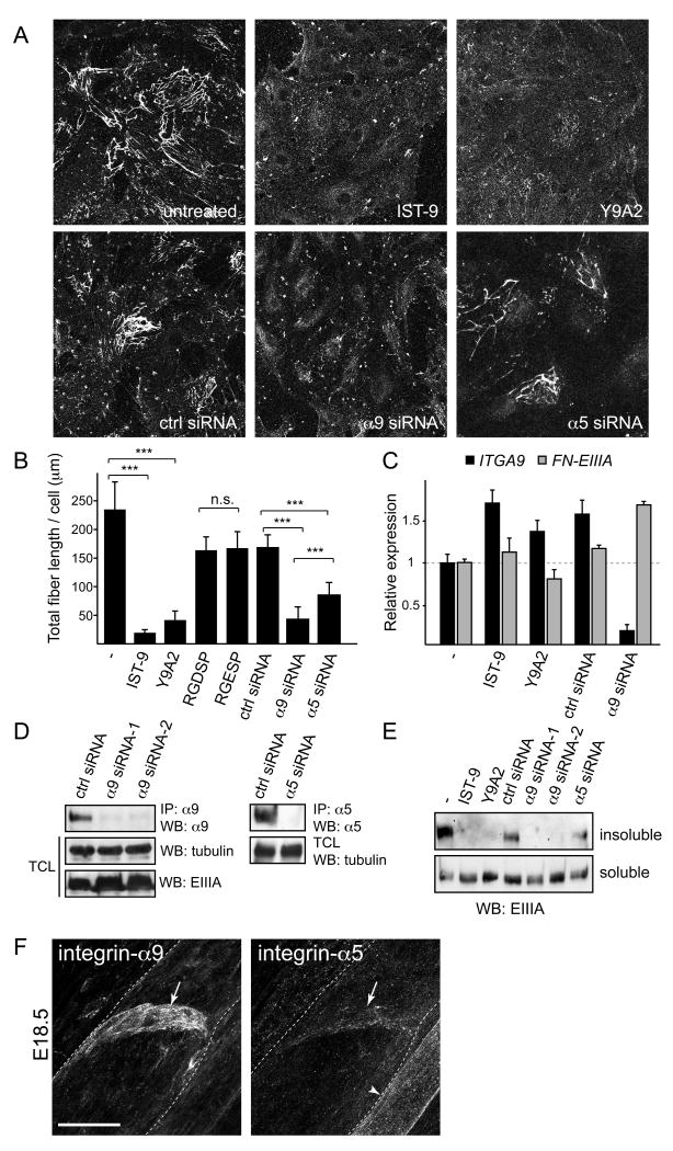
Figure 7. Integrin-α9-EIIIA interaction regulates FN fibril assembly in primary human lymphatic endothelial cells.
(A) FN fibrils in primary human lymphatic endothelial cells (LECs). Integrin-α9-EIIIA interaction was blocked using antibodies against EIIIA (IST-9) or integrin-α9β1 (Y9A2), or siRNA against integrin-α9 or -α5, and stained with EIIIA antibodies.
(B) Quantification of FN fibrillogenesis in the LECs, in which integrin-α9-EIIIA interaction (IST-9, Y9A2, α9 siRNA) or integrin-α5/RGD-dependent integrin interactions (RGDSP peptide, α5 siRNA) were inhibited, in comparison to the control cells (untreated, ctrl siRNA or RGESP peptide). Data represent mean FN-EIIIA fiber length per cell (± s.d) from five randomly chosen view fields in two independent experiments. *** p< 0.003, n.s.= non-significant, p = 0.881 (Student T-test).
(C) qPCR of ITGA9 and FN-EIIIA in human LECs. Data represent mean ± s.d. of triplicates.
(D) siRNA mediated knock-down of integrin expression in primary human LECs. Western blot analysis of immunoprecipitated (IP) cell lysates using integrin-α9 or -α5 antibodies (upper panels). For the loading control, the total cell lysates (TCL) were blotted against α-tubulin and EIIIA.
(E) Conversion of DOC-soluble FN fibrils into insoluble stable matrix. DOC-insoluble (upper panel) and -soluble matrix (lower panel) isolated from the LECs were separated in non-reducing SDS-PAGE and probed for EIIIA.
(F) Immunofluorescent staining of wild type E18 mesenteric vessels using antibodies against integrin-α9 (left panel) and integrin-α5 (right panel). Note low levels of integrin-α5 expression in the valve (arrows) in comparison to strong staining in the blood vessel endothelia (arrowhead).
Scale bar = 20 μm.

