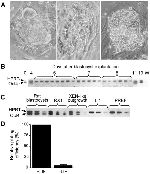
Figure 1. Properties of rat blastocyst outgrowths.
(A) Phase contrast photographs showing stages of WKY rat blastocyst outgrowths kept on mitomycin-treated primary rat embryo fibroblasts (PREF). The outgrowths were initially smooth and compact (left), but converted to XEN morphology (right) ∼10 days after blastocyst plating if not passaged, or a few days later if mechanically disaggregated into smaller clumps. Regardless of when the conversion occurred, it was fast (<24 hours) and went through a stage of intermediate morphology (middle). (B) Loss and re-expression of Oct4 mRNA in WKY rat blastocyst outgrowths. In these experiments, the outgrowths were not passaged and showed compact, smooth morphology before day 10, but XEN morphology thereafter. At the indicated days, the outgrowths were individually harvested for RT-PCR analysis, using rat-specific primers for Oct4 and hypoxanthine phosphoribosyl transferase (Hprt) cDNAs. The Oct4 and Hprt cDNAs were amplified in the same reaction; none of the primers amplified intronless products from genomic DNA (not shown). No amplification was achieved when using mouse-specific primers (not shown). Day 0 = blastocyst; W, water control. (C) Semi-quantitative assessment of Oct4 mRNA level. Rat blastocysts (E4.5, strain WKY), XEN-P line RX1, primary XEN-like blastocyst outgrowths (strain WKY), rat embryo fibroblast line Li 1 (feeder for RX1), and PREF (feeder for primary rat cells) were analyzed for Oct4 and Hprt mRNAs by subjecting 10-fold serial dilutions of the RT reactions to PCR. (D) LIF effect (1,000 u/ml) on the formation of secondary XEN-like cell colonies from primary rat blastocyst outgrowths (WKY). Primary cells were seeded at ∼100–500 cells/well onto feeder line Li 1. 6 independent experiments. Similar results were obtained with rat strain BDIX.

