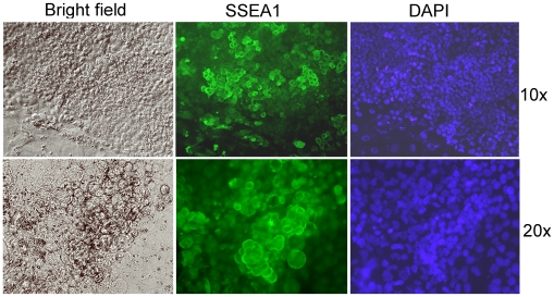
Figure 5. Continued proliferation and preferential accumulation of SSEA1-positive cells in old rat colonies derived from rat XEN-P cells.
Two magnifications of a representative 16-days old colony (line RX1) are shown. Bright field (left), immunofluorescence (middle), and nuclear stain (right). Control omitting primary antibody was negative and is not shown.

