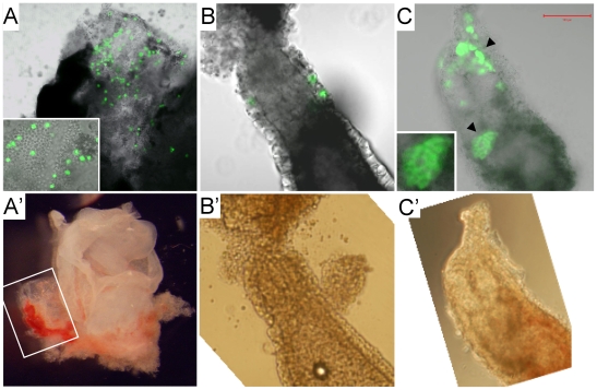
Figure 7. Contributions of rat XEN-P cell lines to postimplantation embryos.
Representative fluorescence (A–C) and bright field (A'–C') photographs demonstrating in vivo contributions of microinjected rat cells to (A, A') parietal yolk sac of an 11.5 dpc rat conceptus (inset showing magnification); (B, B') visceral endoderm of an 8.5 dpc rat conceptus; (C, C') visceral endoderm (arrowheads; one patch magnified in inset) of an ∼7 dpc mouse conceptus. Pregnancy timing is distorted by the embryo manipulations and therefore only approximate.

