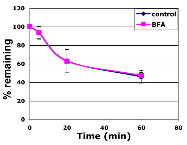
Figure 4. Cell surface Tyrp1 undergoes constitutive recycling.
Melan-a cells that were untreated (blue line) or treated for 60 min with 10 μM BFA (pink line) were incubated with Alexa488-conjugated TA99 on ice followed by incubation for 5 min at 37°C to internalize antibody/antigen complex in the absence or present of BFA. Cells were then incubated (+/- BFA) with anti-Alexa488 quenching antibody or a control non-quenching antibody for the indicated chase times. Remaining fluorescence was quantified by flow cytometry. Mean fluorescence intensity values for quenched samples were divided by values for non-quenched samples at each time point as a measure of % recycled antibody. Values represent mean +/- S.E.M. of three separate experiments each done in duplicate.

