
Figure 6. Pol-III is required for the production of IFN-inducing RNA.
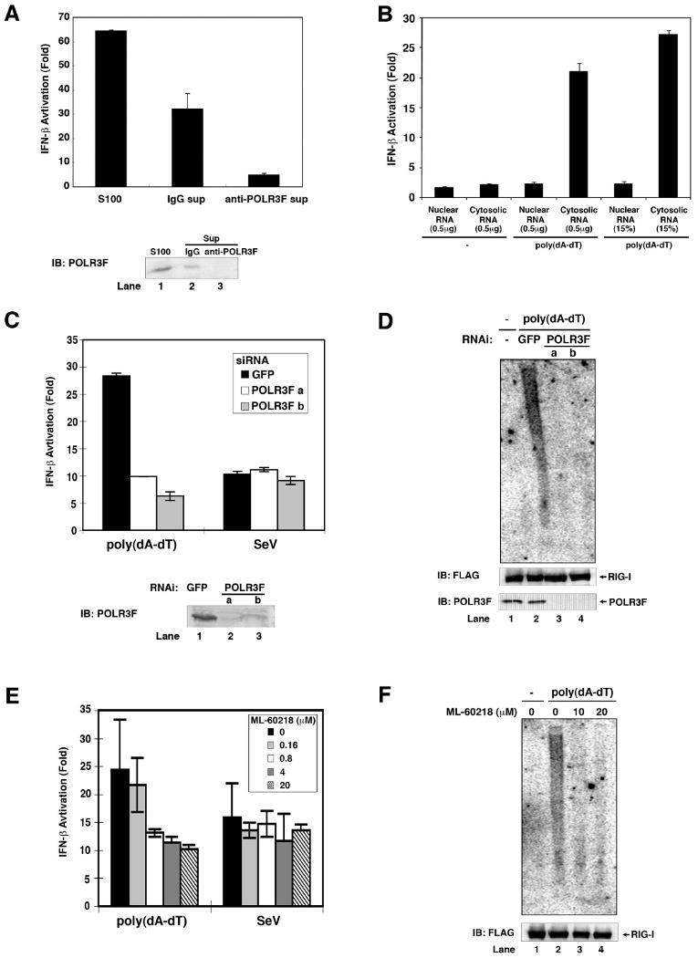
(A) HeLa cell lysate (S100) was immunodepleted five times with a POLR3F specific antibody or control IgG. S100 and the immunodepleted supernatants were incubated with poly(dA-dT), UTP and ATP before RNA was extracted for IFN-β reporter assays (upper panel). The efficiency of immunodepletion was analyzed by immunoblotting with the POLR3F antibody (lower panel). (B) HEK293 cells were transfected with or without poly(dA-dT) for 4 hours. Nuclear and cytosolic lysates were prepared for RNA extraction. 0.5 μg or 15% of nuclear or cytosolic RNAs were transfected into HEK293-IFNβ-luciferase reporter cells to measure IFN-β induction. (C) Two distinct siRNA oligos against POLR3F (a and b) or a control siRNA against GFP were transfected into HEK293 cells. 48 hours after transfection, the cells were transfected with poly(dA-dT) or infected with Sendai virus for 2 hours before RNAs were extracted for IFN-β reporter assays (upper). The efficiency of RNAi was analyzed by immunoblotting with an antibody against POLR3F (lower). (D) HEK293/RIG-I-FLAG stable cells were transfected with siRNA as described in (C), then transfected with poly(dA-dT) for 3 hours. The RIG-I complex was immunoprecipitated using a FLAG antibody, then the RNAs associated with RIG-I were extracted and analyzed by Northern blotting using 32P-labeled poly(A-U) RNA as a probe. The RIG-I in the immunoprecipitates and POLR3F in cell lysates were analyzed by immunoblotting. (E) HEK293 cells were treated with the indicated concentrations of the Pol-III inhibitor ML-60218 for 10 hours before transfection with poly(dA-dT) or infection with Sendai virus for 2 hours. RNAs were extracted for IFN-β reporter assays. (F) HEK293/RIG-I-FLAG stable cells were treated with ML-60218 and then transfected with poly(dA-dT). RNAs associated with the RIG-I complex were analyzed by Northern blotting as described in (D).