
Figure 1. Endothelial cells and mural cells interact.
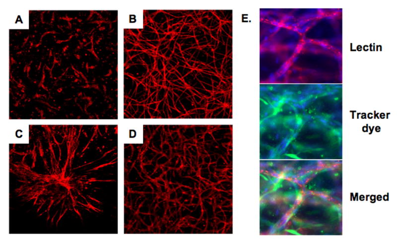
Images of blood vessels formed by endothelial cells (HUVECs) cultured alone (A), or cocultured with human dermal neonatal fibroblasts (HDFNs) (B), human umbilical artery smooth muscle cells (HUASMCs) (C) and human coronary artery smooth muscle cells (HCASMCs) (D) in a 3-dimensional angiogenesis assay. Cells were grown in a collagen matrix for five days, fixed, and stained with TRITC labeled endothelial-specific lectin (red), 100× magnification. (E) Triple labeling with lectin (red), a preloaded CellTracker dye (green) to visualize HDFNs surrounding vessels, and DAPI (blue), 400× magnification.