
Figure 2. Endothelial cells induce NOTCH3 expression in mural cells.
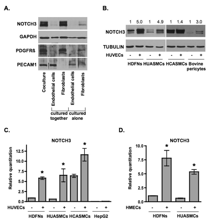
(A) Endothelial cells (HUVECs) and fibroblasts (HDFNs) were cultured alone or cocultured in 3-dimensional angiogenesis assays. After five days, cells were separated by anti-PECAM1-conjugated Dynabeads for Western blot analysis. The purity of the fibroblast and endothelial fractions were evaluated by probing for PDGFRβ as a marker for fibroblasts and PECAM1 as a marker for endothelial cells. (B) HDFNs, HUASMCs, HCASMCs, and bovine pericytes were cultured in 2-dimensions in the presence or absence of HUVECs for 48 hours, separated and subjected to Western analysis. Numbers reflect relative protein expression determined by average pixel intensity from 3 experiments normalized to respective control, P < 0.05. (C) qPCR analysis of NOTCH3 mRNA in HDFNs, HUASMCs, HCASMCs, and HepG2, cultured with or without HUVECs. (D) qPCR analysis of NOTCH3 mRNA in HDFNs and HUASMCs which were cultured alone or cocultured with HMECs. * P < 0.05 compared to control.