
Figure 5. Endothelial-induced NOTCH3 expression requires Notch signaling.
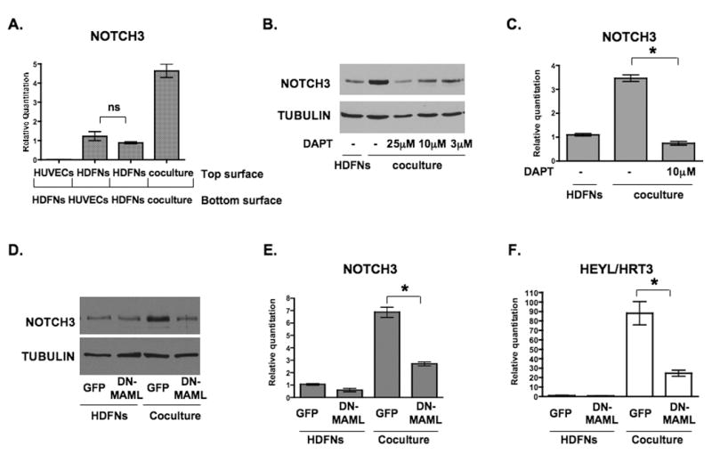
(A) HUVECs and HDFNs were plated on either side of the transwell insert as indicated, cultured for 48 hours, and the cells from the top surface were harvested for qPCR to examine NOTCH3 mRNA expression. (B) Western blot to detect NOTCH3 expression in HDFNs cocultured with HUVECs with varied amounts of the γ-secretase inhibitor (DAPT). Fibroblasts cultured alone were used as a control. (C) qPCR to measure NOTCH3 mRNA expression in HDFNs cocultured with HUVECs in the presence or absence of DAPT. (D-F) HDFNs were lentivirally transduced with GFP or DN-MAML, then cultured alone or cocultured with HUVECs. Fibroblasts were separated from HUVECs for Western analysis to detect NOTCH3 protein (D), or for qPCR to measure NOTCH3 (E) and HEYL/HRT3 (F) mRNA. * P < 0.05; ns, not significant.