
Figure 7. Jagged1 expression is reduced in the absence of Notch3 in vivo.
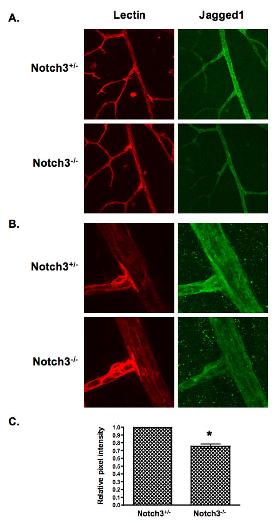
Retinas isolated from Notch3 null (Notch-/-) and heterozygous (Notch+/-) mice at postnatal day 15 were immunostained to detect Jagged1 protein (green). An endothelial-specific lectin (red) was used to highlight the vasculature. Confocal images were taken at (A) 400× and (B) 630× (zoom2) magnification and pixel intensity of Jagged1 staining was quantified (C). Graph represents average Jagged1 expression from 8 null and 8 heterozygous retinas. * P = 0.0004.