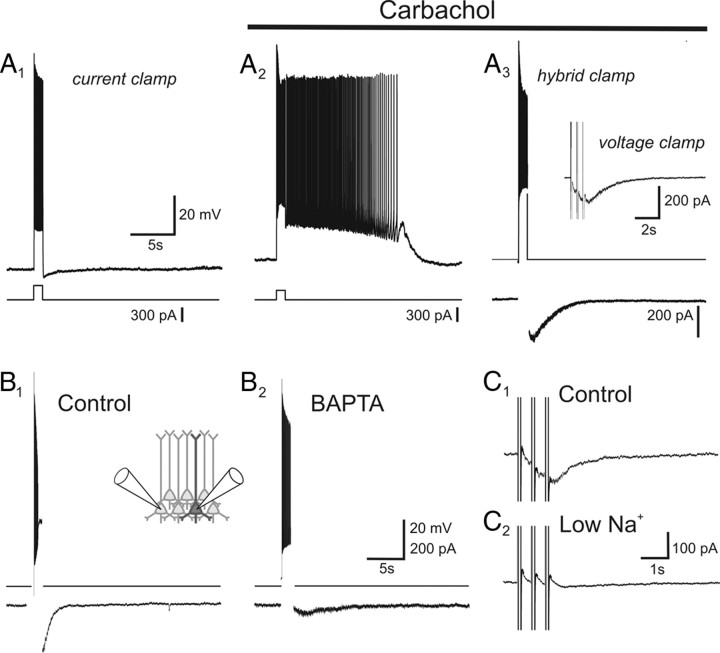
Figure 1.
Muscarinic receptors activate IsADP in organotypic cortical slices. A1, Under basal conditions, a suprathreshold depolarizing pulse delivered to a layer V cortical pyramidal cell of the prefrontal cortex is followed by a slow afterhyperpolarization. A2, Administration of carbachol (30 μm) leads to the replacement of this afterhyperpolarization by an sADP that supports sustained spiking. A3, The current underlying this sADP (IsADP) can be recorded using a hybrid current–voltage-clamp protocol in which it appears as a slow inward aftercurrent following the spike burst. Inset, IsADP can also be triggered by depolarizing steps to 0 mV in voltage clamp. B1, B2, The carbachol-induced IsADP is greatly suppressed when intracellular calcium is buffered near rest using BAPTA. B1 and B2 depict a paired recording from two pyramidal cells in which B1 was recorded using control intracellular solution and B2 using an intracellular solution containing BAPTA to buffer intracellular calcium. C1, C2, Reducing extracellular sodium inhibits IsADP in the control cell depicted in B1 and B2.

