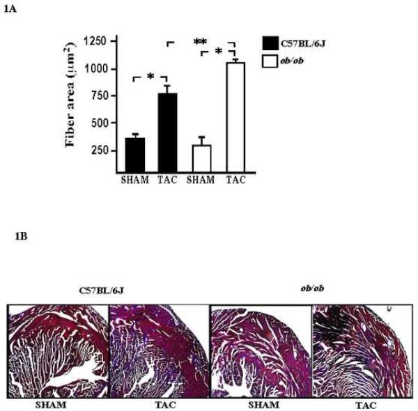
Fig. 1. Morphological changes in wild type and ob/ob mice hearts subjected to TAC.
A, light microscopic analysis was used to determine the myocyte cross sectional area in mice subjected to TAC as compared with those from sham animals in both groups. The increase in myocyte cross sectional area was significant in C57BL/6J and ob/ob mice subjected to TAC. Cross-sectional area was measured using the morphometric system NIH 1.63f. * P values < 0.05 versus sham C57BL6/J; **P < 0.01 versus TAC C57BL6/J (Magnification at 400x). B, collagen deposition in wild type and ob/ob mice subjected to TAC was obtained by the Masson trichrome stain in sections of the left ventricle (Magnification 200x).

