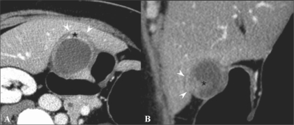
Figure 8 (A,B).
A 54-year-old woman with gallbladder cancer. Axial CT scan (A) shows eccentric wall thickening and a papillary lesion (*) in the fundus of the gallbladder. The fat plane between the liver and the gallbladder seems to be preserved in the axial plane (arrowheads). This lesion was interpretated as T2 on the axial CT image. Oblique coronal MPR image (B) reveals focal hepatic invasion (arrowheads), thus changing the staging and management. Asterisk indicates the papillary lesion

