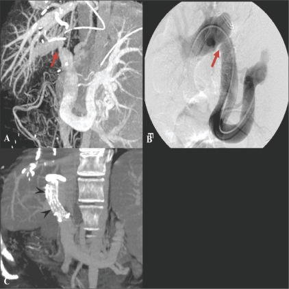
Figure 12 (A-C).
A 45-year-old woman with portal venous stenosis that occurred 8 days after liver transplantation. Coronal oblique MIP image (A) reveals a tight stenosis (arrow) at the portal vein anastomosis. Anterioposterior view of a direct main portal venogram (B) confirms the stenosis (arrow). The stenosis disappeared following metallic stent placement. Follow-up coronal MIP CT scan image (C) shows a patent stent (arrowheads) in the main portal vein

