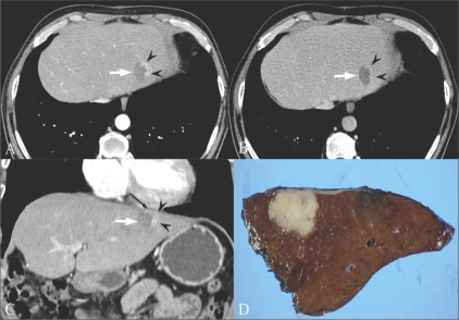
Fig. 14 (A-D).
A 55-year-old man was referred due to marginal recurrence of cholangiocarcinoma at the site of a previous radiofrequency ablation. Arterial phase (A) and portal venous phase (B) axial CT scans show a marginally enhancing lesion (arrowheads in A) with wash-out (arrowheads in B) at the periphery of the previously ablated site (arrow) in the lateral segment of the left lobe of the liver. Initially, repeat radiofrequency ablation was considered for this recurrent mass. However, the coronal MPR image (C) revealed close proximity between the suspected viable tumor (arrowheads) and the inferior pericardium (thin black arrow). To avoid thermal damage to the pericardium and to guarantee a safe and clear margin from the viable tumor, the patient underwent surgery. The lesion was separated from the pericardium and pathologically proven to be a recurrent cholangiocarcinoma. This case demonstrates the role of MPR images as a guidance to determine which procedure is appropriate. MPR : multiplanar reconstruction

