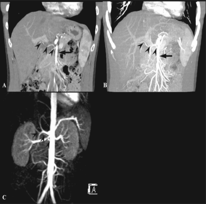
Figure 15 (A-C).
A 35-year-old man with a variation in HA anatomy. Arterial phase MPR (A) and MIP (B) CT scan images show the right HA (arrowheads) arising from the SMA (arrow). MRI (C) also demonstrates a variation in the right HA, Arterial phase MIP MRI also demonstrates the right HA (arrowheads) arising from the SMA (SMA is masked by the aorta). HA: hepatic artery, SMA: superior mesenteric artery

